38 muscle fiber model with labels
Muscle Fiber Model (Altay) Flashcards | Quizlet Muscle fiber model identifications Terms in this set (21) sarcolemma Identify the membrane. endomysium Identify the tissue layer. myofibril Identify the structure. thick myofilament Identify the structure. thin myofilament Identify the structure. neuromuscular junction Identify the connection. axon Identify the structure. axon terminals muscle tissue labeled Muscular system anatomy:muscle fiber with sarcomere model description. Muscle skeletal tissue histology tissues labeled microscope smooth human anatomy slides normal lab physiology medical muscular flashcards biology tejido system. Smooth muscle longitudinal section40x light micrograph high-res stock muscle tissue labeled
Muscle Fibers: Anatomy, Function, and More - Healthline Each muscle fiber contains smaller units made up of repeating thick and thin filaments. This causes the muscle tissue to be striated, or have a striped appearance. Skeletal muscle fibers are...

Muscle fiber model with labels
anatomy labeling muscle anatomy kenhub constrictor muscles ridge medius muscle pharynx pharyngeal musculus middle passavant aspects clinical. Gross Anatomy Of The Muscular System Review Sheet boundbobskryptis.blogspot.com. anatomy. Abdominal Muscle Labeling Quiz . abdominal muscle quiz. Skeletal Muscle Fiber Model . muscle ... Muscle Models | Muscle Figures | Musculature Models - 3B Scientific Explore the muscles in 3D using the MICROanatomy™ Muscle Fiber. A single striated muscle fiber is magnified 10,000 times along with its neuromuscular end plate. Break down the muscular anatomy of the leg and hip (9-parts) and the muscles of the arm... 3/4 Life-Size Dual Sex Human Muscle Model on Metal Stand, 45-part - 3B Smart Anatomy $ 9,003.00 Muscle Fiber Model: Motor Neuron, Myeline Sheath, Node of Ranvier ... Muscle Fiber Model: Motor Neuron, Myeline Sheath, Node of Ranvier, Synaptic Terminal, Synaptic Cleft, Endomysium, Sarcolemma, Nuclei, Mitochondria, T-tubules, Sarcoplasmic Reticulum, Myofibrils Find this Pin and more on Strength Training Benefits by Great Bones. Motor Neuron Cell Model Musculoskeletal System Anatomy Models Muscular System
Muscle fiber model with labels. Muscle Fiber Model #1 - Ohio University - Anatomy & Physiology Muscle Fiber Model #1 - Ohio University - Anatomy & Physiology Muscles Labeling - The Biology Corner The activity linked below is a drag and drop activity for students to practice labeling the muscles, there are 6 slides showing images of muscles and fibers and the connective tissue surrounding the fibers (endomysium, perimysium, epimysium). Google Slides Key (TpT) Prev Article Next Article Solved Lab 9: Muscle Tissue and axial muscle Exercise 1. | Chegg.com Identify and list a function of each labeled item 1-8) on the models Below. These are the same model at different angles. These are models of ONE muscle fiber or one skeletal muscle cell. Use the following terms to help you label: Endomysium, sarcolemma, myofibril, sarcoplasmic reticulum, myofilaments, motor neuron, T-tubule, nucleus. Muscle fiber model Quiz - PurposeGames.com This is an online quiz called Muscle fiber model. There is a printable worksheet available for download here so you can take the quiz with pen and paper. Your Skills & Rank. Total Points. 0. Get started! Today's Rank--0. Today 's Points. One of us! Game Points. 13. You need to get 100% to score the 13 points available.
Muscular System Labeled Diagram Pictures, Images and Stock Photos Rear human structure model with medical titles for healthcare study handout vector illustration. Parts location example. muscular system labeled diagram stock illustrations ... scheme. Labeled medical infographic. Motor neuron and muscle cell structure closeup. Diagram with myofibril and muscle fibers. muscular system labeled diagram stock ... labeled muscle for anatomy stomach histology tissue tract digestive gastrointestinal system human anatomy slides embryology labeled upper fundus epithelial intestine muscularis cardias edu del. MUSCULAR SYSTEM ANATOMY:Muscle Fiber With Neuromuscular Junction Model . muscle junction neuromuscular fiber anatomy muscular system. Untitled Document [bio ... cardiac muscle cell labeled cardiac muscle cell labeled Muscular system anatomy:muscle fiber with sarcomere model description. Muscle cardiac histology heart tissue cell cells slides anatomy human cardiovascular system embryology medical science labeled microscope physiology edu slide. Cardiac otot striated skeletal fibres jaringan jantung ocr ucl cardiac muscle cell labeled Sarcomere Model | Muscle Fiber Model | Skeletal Muscle | MICROanatomy ... This micro-anatomy model magnifies the anatomy of the human muscle fiber approximately 10,000 times. This muscle model illustrates a section of a skeletal muscle fiber and its neuromuscular end plate. The muscle fiber is the basic element of the diagonally striped skeletal muscle. You've never seen a muscle fiber in this way!
PDF MUSCLE MODEL ACTIVITY GUIDE - Field Museum of Natural History muscle model / The Machine inside: BioMechanics activity guide For stud E nt ACTIVITY - Exploring Muscle Size Most animals, including humans, are born with the exact number of muscle fibers they will have their entire lives. This means they cannot increase their density, but they can change the size of muscle fibers. Explore how fiber size ... Muscle Anatomy Models Anatomy Now is the leading provider of 3D human anatomical muscle system models, charts, and replica models for the medical profession and education of patients. There are three distinct types of muscles: skeletal muscles, cardiac or heart muscles, and smooth muscles. ... Microanatomy Muscle Fiber #B60. Retail: $290.00 Our Price: $265.00. Human ... Pin page - Pinterest This micro-anatomy model magnifies the anatomy of the human muscle fiber approximately 10,000 times. This muscle model illustrates a section of a skeletal muscle fiber and its neuromuscular end plate. The muscle fiber is the basic element of the diagonally striped skeletal muscle. You've never seen a muscle fiber in this way! This high quality ... Skeletal Muscle Histology Slide Identification and Labeled Diagram ... The skeletal muscle fibers are elongated, cylindrical and multinucleated cells whose length may vary in different animals. In this short guide, you will get a basic concept of skeletal muscle histology from the real slide and labeled diagram. You will also get the identification points of skeletal muscle histology slide with a little description here in this guide.
V Ling: Art Center Summer Show UPDATE
anatomy labeled muscle fiber - Microsoft Artery vein histology muscle anatomy muscular smooth arteries microanatomy layers tissue vessel slide blood elastic exam type identify vs vessels. The skeletal muscle fiber stock vector. Microscopic structure of skeletal muscle by dr. s. n. singh anatomy labeled muscle fiber

rfumsphysiology / Muscle Physiology I
Muscle Model - an overview | ScienceDirect Topics The muscle models have been used in the exoskeleton control schemes. Unlike the dynamic model, the muscle model predicts the muscle forces deployed by the muscles of the human limb joint as a function of muscle neural activities and the joint kinematics (Anam and Al-Jumaily, 2012). The input is the EMG signals and the output is force estimation.

Ben Smith: Textures (feels good)
muscle fiber model labeling Diagram | Quizlet muscle fiber model labeling STUDY Learn Flashcards Write Spell Test PLAY Match Gravity Created by crlavenuePLUS Terms in this set (7) transverse tubule sarcoplasmic reticulum triad sarcolemma myofibril consists of actin (thin) & myosin (thick) fibrils sarcomere nucleus Subjects Arts and Humanities Languages Math Science Social Science Other
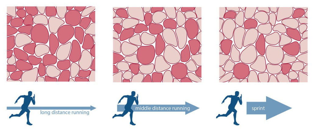
Skeletal Muscle Fiber Type: Influence on Contractile and Metabolic Properties
Skeletal Muscle Fiber Labeling - Printable This is a free printable worksheet in PDF format and holds a printable version of the quiz Skeletal Muscle Fiber Labeling. By printing out this quiz and taking it with pen and paper creates for a good variation to only playing it online.

Gantungan kunci karet, Frosted, Fiberglass, Digital printing, Emboss, Kaca patri, Seni kaca ...
Skeletal Muscle Fiber - GetBodySmart Skeletal Muscle Fiber. Skeletal muscles are a type of striated muscle. They are attached to the bones of the skeleton by tendons. Skeletal muscle fibers have a striated (striped) appearance because they are made up of smaller units called sarcomeres that run parallel to each other.. A sarcomere is the smallest functional unit of skeletal muscle tissue, and each sarcomere has thick and thin ...

Muscle fiber model
General Anatomy of Skeletal Muscle Fibers | GetBodySmart Skeletal Muscle Fiber Location and Arrangement. are located inside muscles, where they are organized into bundles called […] Internal Anatomy of Skeletal Muscle Fibers. An interactive quiz about the internal anatomy of skeletal muscle fibers, featuring illustrations-based multiple choice questions.

357 best Nursing School and Study Guides images on Pinterest
Cardiac Muscle Fiber Model - Spectrum Scientifics Over $1000 Description This block model depicts a cardiac muscle fiber that has been greatly enlarged to represent significant anatomical features such as the intercalated discs. Also includes detailed representations of sarcolemma, T-tubules, myofilaments, nucleus, and mitochondria. Mounted on a base. Dimensions: 15.75" x 10.25" x 7.5"

35 Label The Structures Of A Skeletal Muscle - Labels Design Ideas 2020
Muscle Fiber Micro Anatomy Models - GTSimulators.com Muscle Fiber Micro Anatomy Models Sort by Best Selling Grid List Complete Sarcomere Model (0779-00) Reviews Item # DGAP79 U.S. Contiguous States Only $1,150.00 $1,058.00 Add to cart 3B MICROanatomy Muscle Fiber Model - 10,000 times magnified Reviews Item # B60 $339.00 $312.00 Add to cart Free U.S. Shipping

Chapter 9: Muscles and Muscles Tissue | Anatomy and Physiology | Pinterest | Muscle tissue ...
muscle fiber diagram labeled Show The Different Between The Three Types Of Muscle Fibers With brainly.in muscles types muscle skeletal fibers three different diagram smooth striated cell body system muscular type involuntary human cells brainly heart Protein actin diagram structure muscle contraction labeled illustration vector illustrations skeletal istockphoto clip.

Neurolemmocyte On Skeletal Muscle Model - Human Anatomy - GUWS Medical
Skeletal Muscle Fiber Model - Myofibrils - YouTube This video was produced to help students of human anatomy at Modesto Junior College study our anatomical models.

This photo shows a model of an osteon. It points out the blood vessels and shows the different ...
SAC A&P Model Key - Muscular System Muscular System. M1 - Muscled Arm. M2 - Muscle Leg. M3 - Female Muscle Figure. M4 - Microanatomy Muscle Fiber. M5 - Muscle Figure.

Sarcomere model - YouTube
Muscle Fiber Model: Motor Neuron, Myeline Sheath, Node of Ranvier ... Muscle Fiber Model: Motor Neuron, Myeline Sheath, Node of Ranvier, Synaptic Terminal, Synaptic Cleft, Endomysium, Sarcolemma, Nuclei, Mitochondria, T-tubules, Sarcoplasmic Reticulum, Myofibrils Find this Pin and more on Strength Training Benefits by Great Bones. Motor Neuron Cell Model Musculoskeletal System Anatomy Models Muscular System
Types Muscle Tissue Skeletal Muscle Smooth Stock Illustration 138354809 - Shutterstock
Muscle Models | Muscle Figures | Musculature Models - 3B Scientific Explore the muscles in 3D using the MICROanatomy™ Muscle Fiber. A single striated muscle fiber is magnified 10,000 times along with its neuromuscular end plate. Break down the muscular anatomy of the leg and hip (9-parts) and the muscles of the arm... 3/4 Life-Size Dual Sex Human Muscle Model on Metal Stand, 45-part - 3B Smart Anatomy $ 9,003.00

V Ling: 08.10
anatomy labeling muscle anatomy kenhub constrictor muscles ridge medius muscle pharynx pharyngeal musculus middle passavant aspects clinical. Gross Anatomy Of The Muscular System Review Sheet boundbobskryptis.blogspot.com. anatomy. Abdominal Muscle Labeling Quiz . abdominal muscle quiz. Skeletal Muscle Fiber Model . muscle ...

Replika, Taman, Meubel, Jualan, Franchise, Pembantu, Pegawai, Mahasiswi, Guru, Gordyn, Jok ...

Ch. 9 Muscles and Muscle Tissue Flashcards | Easy Notecards
10.06.20 - Types of Muscle Fibers - Amanda Jackson Whitney | Library | Formative
Post a Comment for "38 muscle fiber model with labels"