45 microscope images with labels
Histology and Microscope Slide Labels - emsdiasum.com Microscope Slide Labels. These specialty Microscope Slide Labels and matching End Labels are available in standard (thin) or pathology (tissue high) thickness, and square or round corner (RC). Permanent adhesive holds labels in place during use and long-term storage. Sheet Form Size is 5¼" x 8". Prices are per thousand labels. Slide Label Microscopic Image Annotation and - Columbia University Starting with the expert labeling of a few cells according to some predefined phenotypes, the system learns to infer the phenotype classes of unlabeled cells on the microscopic images. The learning is done in a semi-supervised manner that both the labeled and unlabeled data are utilized. Given the predicted phenotype label for the cells, image-level relevance scores are also computed.
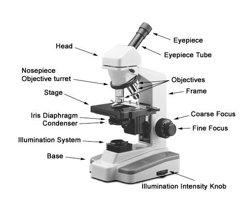
Parts of a microscope with functions and labeled diagram - Microbe Notes Q. Differentiate between a condenser and an Abbe condenser. Ans. Condensers are lenses that are used to collect and focus light from the illuminator into the specimen. They are found under the stage next to the diaphragm of the microscope. They play a major role in ensuring clear sharp images are produced with a high magnification of 400X and above.

Microscope images with labels
Simple Microscope - Parts, Functions, Diagram and Labelling Simple Microscope - Parts, Functions, Diagram and Labelling A microscope is one of the commonly used equipment in a laboratory setting. A microscope is an optical instrument used to magnify an image of a tiny object; objects that are not visible to the human eyes. Table of Contents The common types of microscopes are: What is a Simple microscope? PDF Label parts of the Microscope Label parts of the Microscope: . Created Date: 20150715115425Z Microscope Labeled Pictures, Images and Stock Photos Browse 48 microscope labeled stock photos and images available, or start a new search to explore more stock photos and images. Newest results. Fluorescent Imaging immunofluorescence of cancer cells growing in 2D with nuclei in blue, cytoplasm in red and DNA damage foci in green microscope labeled stock pictures, royalty-free photos & images.
Microscope images with labels. Parts of a Simple Microscope - Labeled (with diagrams) image 5: A modern simple microscope with the different parts labeled. image source: laboratoryinfo.com The optical parts of a simple microscope are centered on the specimen - lighting, and magnification. › microscopy › intSmart Microscope for Lab Routine and Research - ZEISS Your smart microscope from ZEISS lets you assign microscope images with the correct scaling information to barcode-labeled samples. Just use your Axiolab 5 or Axioscope 5 microscope with Windows PC or iPad, connect a barcode reader to your Axiocam 208 color microscope camera and you’re good to go. A Study of the Microscope and its Functions With a Labeled Diagram A Study of the Microscope and its Functions With a Labeled Diagram To better understand the structure and function of a microscope, we need to take a look at the labeled microscope diagrams of the compound and electron microscope. These diagrams clearly explain the functioning of the microscopes along with their respective parts. Microscope Parts and Functions The specimen is placed on the glass and a cover slip is placed over the specimen. This allows the slide to be easily inserted or removed from the microscope. It also allows the specimen to be labeled, transported, and stored without damage. Stage: The flat platform where the slide is placed.
› dinocaptureDinoCapture 2.0: Microscope Imaging Software | Dino-Lite Dino-Lite USB microscope cameras include DinoCapture 2.0, the powerful yet easy to use microscope imaging software for Windows. DinoCapture is a professional microscope imaging software that was made for users of all levels, including basic features from image viewing and capture, measurement with calibration, to advanced features such as geotags and edge detection. Labeling the Parts of the Microscope | Microscope World Resources Labeling the Parts of the Microscope This activity has been designed for use in homes and schools. Each microscope layout (both blank and the version with answers) are available as PDF downloads. You can view a more in-depth review of each part of the microscope here. Download the Label the Parts of the Microscope PDF printable version here. Labeling the Parts of the Microscope | Microscope activity, Science ... Jan 13, 2016 - Free worksheets for labeling parts of the microscope including a worksheet that is blank and one with answers. Learning to segment microscopy images with lazy labels In this paper, we introduce a deep convolutional neural network for microscopy image segmentation. Annotation issues are circumvented by letting the network being trainable on coarse labels combined with only a very small number of images with pixel-wise annotations. We call this new labelling strategy `lazy' labels.
rsscience.com › stereo-microscopeParts of Stereo Microscope (Dissecting microscope) - Rs' Science Unlike a compound microscope that offers a flat image, stereo microscopes give the viewer a 3-dimensional image that you can see the texture of a larger specimen. [In this image] Examples of Stereo & Dissecting microscopes. Major microscope brands (Zeiss, Olympus, Nikon, Amscope, Omano, Leica …) all produce stereomicroscopes. Labelled Diagram Of A Light Microscope | Products & Suppliers ... Acoustic and ultrasonic microscopes use sound waves to create images of the sample. Compound microscopes use a single light path. These types of ... (LaB6) cathodes, 46, 293 Laser confocal fluorescence microscope, 40 Lectin labels , 38, 51, 175 Lens aberrations, 30-31 Light microscopy (LM), 27, 29, 89, 153, 174, 176-177 colloidal gold ... › cemf › whatisemWhat is Electron Microscopy? - UMASS Medical School Conventional scanning electron microscopy depends on the emission of secondary electrons from the surface of a specimen. Because of its great depth of focus, a scanning electron microscope is the EM analog of a stereo light microscope. It provides detailed images of the surfaces of cells and whole organisms that are not possible by TEM. Microscope Stock Photos, Pictures & Royalty-Free Images - iStock Microscope. Microscope.This royalty free vector illustration features the main icon on both white and black backgrounds. The image is black and white and had the background rendered with the main icon. The illustration is simple yet very conceptual. Science line icons.
31 Compound Microscope With Label - Labels For Your Ideas
Learning to Segment Microscopy Images with Lazy Labels Multi-task learning for image segmentation with lazy labels. The figure uses Scanning Electron Microscopy (SEM) images of food microstructures as an example and demonstrates a segmentation problem of three classes, namely air bubbles (green), ice crystals (red) and background respectively.
Print Microbiology Lab 2 (Microscopy, Gram stain, intro to enterotube II) flashcards | Easy ...
› products › microscopeLAS X Industry Microscope software for Industry | Products ... Create a single sharp image by capturing a stack of images at different focus positions and combining them automatically into an Extended Depth of Focus (EDOF) image. LAS X Extended Depth of Field: Create sharp 2D images from several partially in-focus images. In connection with the 3D Surface Viewer, creation of 3D images is also possible.
1.1 Labelling Microscope - Labelled diagram
› microscopy › intZEISS Axiocam Microscope Cameras for Science and Research For easy documentation, some digital cameras can record images either completely without a computer, or through a connected PC, laptop or iPad running the ZEISS Labscope software. All our microscope cameras are fully supported in our ZEN software environment, offering fast live image display and easy-to-use user interface.

Flares into Darkness: Insect faces
Drawing Of A Microscope And Label - Warehouse of Ideas The optical parts of the microscope are used to view, magnify, and produce an image from a specimen placed on a slide. ... Learn how to draw microscope and label pictures using these outlines or print just for coloring. Source: ugarevwesi.blogspot.com. Supports and stabilizes the microscope. Optical parts of a microscope and their functions.

Stacey Kalkowski's Art Journal: Pollen Spores and Asteroids
Fluorescence Microscopy - Explanation and Labelled Images The samples are labeled with fluorophore where they absorb the high-intensity light from the source and emit a lower energy light of longer wavelength. The resulting fluorescent light is then separated from the surrounding radiation with filters, allowing the observer to see only the fluorescing material.

Amoeba proteus 400x's | Nucleus and pseudopods clear | Flickr - Photo Sharing!
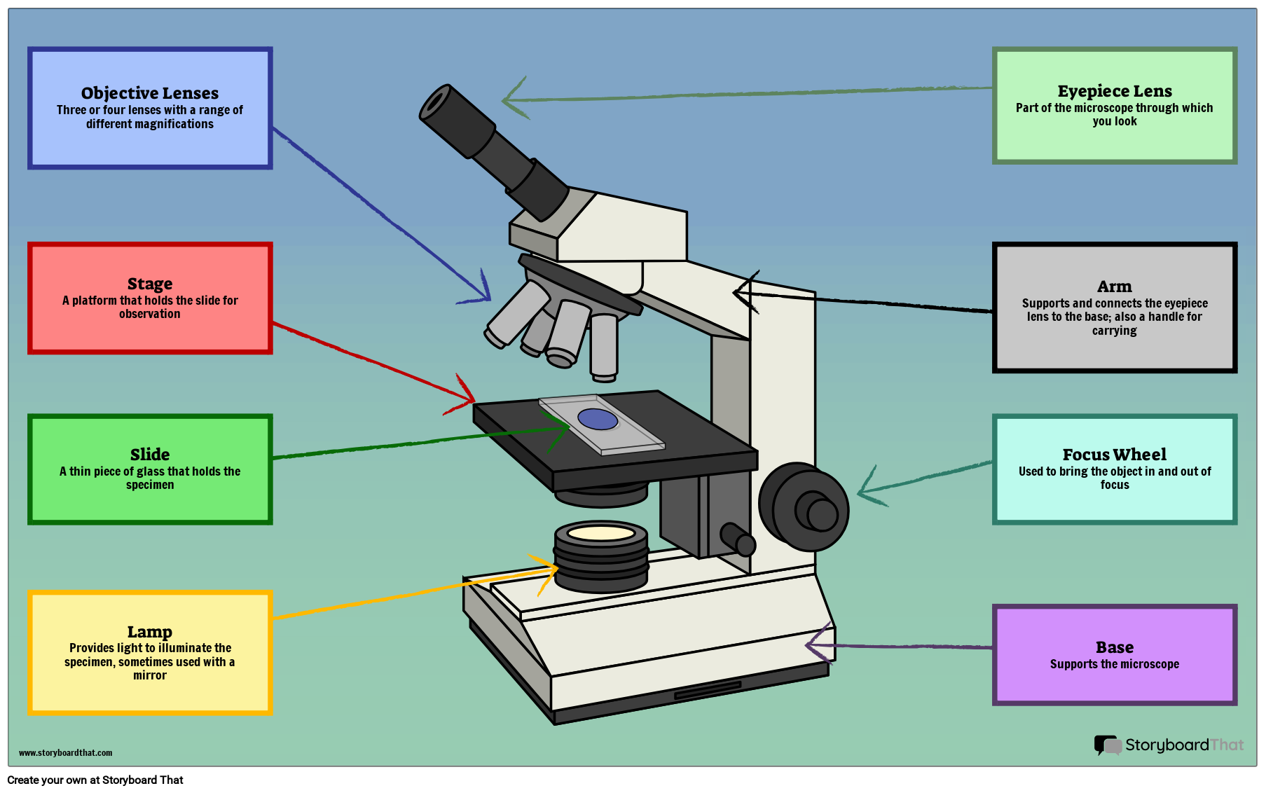
Label the microscope — Science Learning Hub All microscopes share features in common. In this interactive, you can label the different parts of a microscope. Use this with the Microscope parts activity to help students identify and label the main parts of a microscope and then describe their functions. Drag and drop the text labels onto the microscope diagram. If you want to redo an answer, click on the box and the answer will go back to the top so you can move it to another box.

31 Label And Color The Parts Of Both Microscopes - Labels For You
microscope picture with labels - Compound Light Microscope... View microscope picture with labels from BIOL 1005Y at Yeshiva University. Compound Light Microscope ocular (eyepiece) revolving nosepiece objectives coarse adjustment knob mechanical stage fine

Microscope labeling
Electron Microscopy Images - Dartmouth Transmission electron microscope image of a thin section cut through the bronchiolar epithelium of the lung (mouse), which consists of ciliated cells and non-ciliated cells. Image shows the ciliary microtubules in transverse and oblique section. In the cell apex are the basal bodies that are the anchoring sites for the cilia.

Beyond the Human Eye: A tiny aquatic worm that clones itself
Parts of the Microscope with Labeling (also Free Printouts) Microscopes are specially created to magnify the image of the subject being studied. This exercise is created to be used in homes and schools. the microscope layout, including the blank and answered versions are available as pdf downloads. Click to Download : Label the Parts of the Microscope (A4) PDF print version.

Exploration of the Human Spinal Cord
Compound Microscope Parts - Labeled Diagram and their Functions The eyepiece (or ocular lens) is the lens part at the top of a microscope that the viewer looks through. The standard eyepiece has a magnification of 10x. You may exchange with an optional eyepiece ranging from 5x - 30x. [In this figure] The structure inside an eyepiece. The current design of the eyepiece is no longer a single convex lens.

labels of a compound microscope microscope boxed - Top Label Maker
Amazing 27 Things Under The Microscope With Diagrams - Microbe Notes Skeletal muscle under the microscope 40X magnification 100X magnification 400X magnification 20. Skin under the microscope 21. Snowflake under the microscope 22. Sperm under the microscope Direct observation Observation after staining 23. Spirogyra under the microscope 24. Virus under the microscope Fluorescence microscope
31 Picture Of Microscope With Label - Labels Database 2020
› newgrouppage9Educational Atomic Force Microscope (AFM) - Thorlabs Nov 05, 2021 · The images below were all taken using Thorlabs' Educational Atomic Force Microscope (AFM). Of the samples shown below, the microstructure sample and blu-ray disc are included with the kit. If you would like to share your own AFM images, you can submit them to techsupport@thorlabs.com and we'll consider them for addition to the image gallery.

Microscopy Flipped Home Learning - Lessons - Tes Teach
Microscope Labeling - The Biology Corner The google slides shown below have the same microscope image with the labels for students to copy. I often spend the first day walking students through the steps and having them look at a single slide as we do the steps. Students are often very enthusiastic about using microscopes and will try to start with the high power objective.

Onion Cell, 400X | Sue Bachus | Flickr
Polarizing Microscope Image Gallery | Science Lab - Leica Microsystems Images recorded with a DM4 P microscope using transmitted light, 20x N Plan DS (dispersion staining) objective, and polarizers. This image shows the typical magenta-blue dispersion color of chrysotile in an E-W orientation. The medium has a refractive index of 1.553.
Label a microscope - Teaching resources
Compound Microscope with labels Stock Vector | Adobe Stock Download Compound Microscope with labels Stock Vector and explore similar vectors at Adobe Stock. Adobe Stock. Photos Illustrations Vectors Videos Audio Templates Free Premium Editorial Fonts. ... Get 10 free Adobe Stock images. Start now. Get 10 free images. Unlock 200M+ assets in our full collection.

Skin (Integumentary System)
Microscope picture label Flashcards | Quizlet Microscope picture label Flashcards | Quizlet Microscope picture label STUDY Flashcards Learn Write Spell Test PLAY Match Gravity Created by kfire Terms in this set (12) Arm What is the part labelled C? Base What is the part labelled D? Body tube What is the part labelled B? Ocular lens What is the part labelled A? Illuminator

Microscope Picture To Label - Micropedia
300+ Free Microscope & Laboratory Images - Pixabay Find your perfect microscope image. Free pictures to download and use in your next project. 189 37. analysis biochemistry. 335 71. analysis biochemistry. 334 96. microscope slide. 725 186.
Post a Comment for "45 microscope images with labels"